This series of illustrations visually describe the different types of F.G.C (Female Genital Cutting; also know as Female Genital Mutilation).
Each illustration showcases the increasing severity or level of cut. With each image is a description of what has been done in each procedure.
The works here were completed in collaboration with The Sauti Yetu Foundation.
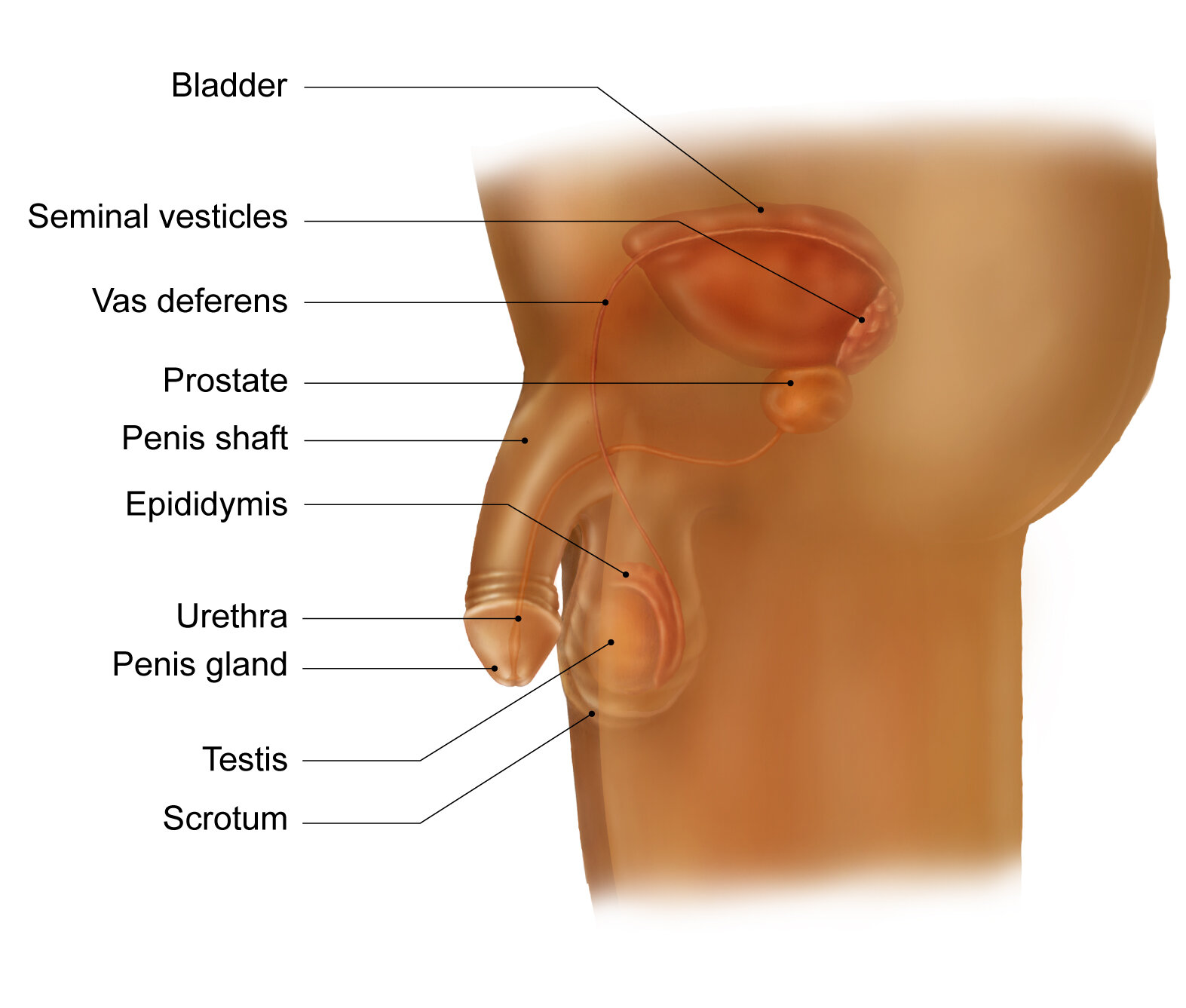
Pre-adolescent Female genital anatomy & Male external and partial internal genital anatomy


1A
Clitoridectomy 1: Removal of Clitois only.
Scar tissue is formed by the remaining clitoris hood

1B
Clitoridectomy 2: Removal of both Clitois gland and clitoris hood
Remaining skin sutured together, creating new scar tissue

2A
Combination of Clitoridectomy 2 and excising of Labia minora.

2B
Combination of Clitoridectomy 2 and excising of both Labia minora and Labia majora.
Scarring between both lips of labia.

3
Complete removal and apposition of skin both lips of labia as well as the complete removal of the structure of the clitoris, resulting in the forming of a cutaneous bridge scar tissue.
The opening of the Urethra is closed by the cutaneous bridge
A new vagina opening is created as a result of the cutaneous bridge